What is subluxation of tooth?
March 28, 2023

The impact of a traumatic injury to the face have an adverse effect on the teeth. Depending on the type of impact, direction of the impact, resilience of the tooth structure and shape of the tooth structure, the tissues surrounding the teeth are also affected. One such type of trauma to the tooth is called subluxation.
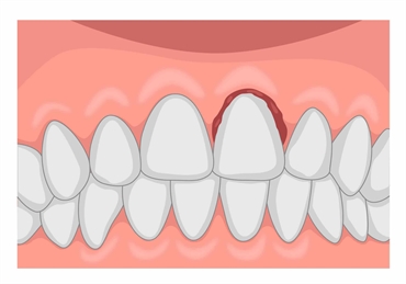
Subluxation is when the tooth-supporting structures are injured, leading to an abnormal loosening of the tooth inside the socket. However, there is no displacement of the tooth following this injury. Alveolar bone, periodontal ligament, cementum and the gingival tissues are the four tissues surrounding the tooth that are affected in subluxation injury of the tooth.
Clinical Findings in subluxaion
When a patient presents with a subluxation injury, a routine examination reveals that the affected tooth has become tender to touch.
There is tooth is mobile without any displacement.
Bleeding from the gingival tissues may also be seen.
The impact of the tooth injury might be felt by the deeper tissues within the tooth, i.e., the pulp. This soft tissue is responsible for the tooth's blood and nervous supply.
Radiographic findings in tooth subluxation
A radiograph is routinely advised in such cases in order to observe the extent of the injury and if any additional internal injuries are seen. Depending on the site involved, the dental surgeon might advise more than one type of radiograph for accurate diagnosis.
Treatment for a subluxated tooth
If the mobility of the tooth is minimal, then physiologic defense mechanisms may be able to rectify the condition without any intervention. However, this minimal level of mobility should be diagnosed only by the experienced clinician and not by the patient. In cases of distinct mobility, the tooth is splinted with a passive and flexible splint. The procedure involves bonding a splint to the affected tooth and the adjacent teeth with biocompatible luting cements. These do not hamper any normal oral functions like speech or mastication.
The patient is kept on a follow-up for at least a year. The usual protocol of follow-up is done at 2 weeks, 12 weeks, 6 months and 1 year.
Prognosis
In most cases, the condition has a favorable prognosis. The tooth is usually asymptomatic after 1 year. The removal of the splint must only be after the dental surgeon satisfactorily ensures the radiographic success of the tooth and its surrounding structure.
The least desired outcomes involves pain, infection and necrosis of the affected tooth. Additionally, the tooth's roots might start resorbing owing to the accelerating infection. In such cases, root canal treatment must be started immediately. If needed, the dental surgeon might also place a calcium-hydroxide-based paste or an antibiotic paste as a medicate agent within them root canals to halt the resorptive process.
Subluxation in a young permanent tooth
Subluxation to a permanent tooth wose roots are yet to be formed requires thorough observation during follow-ups. Following the injury if the apex of the tooth's root or roots is incomplete, then the patient is monitored radiographically in every follow-up. During the recalls, if the dental surgeon feels that there is no progress in the root Alex's closure, then surgical procedures to seal the apex of the teeth are initiated. These are also linked to root canal therapies but are more technically sensitive.
Subluxation in milk teeth
Subluxation of primary teeth is similar in clinical appearance. However, follow-up protocols and treatment outcomes differ as the health of the underlying erupting permanent tooth is also taken into account. Immediate treatment involves advising home remedies which involves cleaning the gingival tissues with 0.2% chlorhexidine and ensuring that no unnecessary trauma or traction is imposed on the affected tooth. The tooth is initially observed at 1 week and 2 months and later at an annual basis until the tooth exfoliates.
During these recalls if the tooth becomes discolored or shows signs of infection and necrosis, then depending on the length of the root, the status of the permanent successor and the extent of apical infection, a treatment plan is formulated. In most cases, the tooth is either saved by a pulpectomy procedure, or it is extracted. In cases of extraction, the socket space is preserved by an appliance called a space maintainer. This allows the preservation of the path of the erupting permanent successor.
Subluxation of the tooth should thus be reported to the dental surgeon at the earliest. The key to prognosis in these cases highly depends on how regular the patient is in his/her follow-up visits.

