What does MIH mean in dentistry
September 29, 2022

MIH in dentistry stands for Molar-Incisor Hypomineralization. This refers to a condition of a selected teeth that was first recognized two decades ago. It is a defect of the surface layer of the teeth that becomes weakened and eventually becomes the basis for carious attacks and broken teeth.
Causes of MIH (Molar-Incisor Hypomineralization)
MIH occurs because of improper development of the tooth that can be attributed to more than one reason.
There is no definitive mechanism that has been established to understand why MIH occurs. Some of the possible reasons for the development of hypomineralised teeth are listed below.
During the last trimester, if the mother suffers from chronic illness or has been on prolonged antibiotics, then it might hinder the with the tooth development of the infant.
Dioxins are chemicals that are also environmental pollutants. Expecting mothers who reside in areas with high dioxin levels get exposed to this pollutant which mixes with the breast milk. Thus, feeding an infant with breast milk that has high content of dioxins can also demineralize the layers of developing teeth.
Complications during child birth can also cause anatomical defrcts that subsequently hamper tooth development in the child.
A child with long-term respiratory conditions is at risk of having MIH.
Children with disorders that affect the metabolism of calcium and phosphate in the body are more likely to have MIH.
Reduced weight and oxygen starvation are other causes that can lead to MIH.
Diagnosis
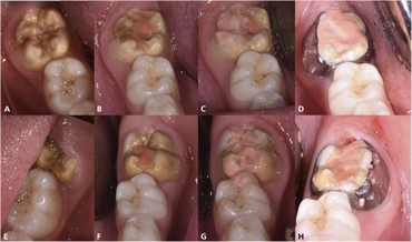
The diagnosis of MIH depends on the affected teeth and the pattern in which they are hypomineralized. Spot lesions more than 1 mm in diameter and of whitish, yellowish or brownish opacities are commonly seen. The incisors and the molars are commonly affected. In more severe cases, the hypomineralization can spread to other teeth.
Patient can present with teeth that are gradually breaking or whose layers are gradually chipping off. They also have a high dentinal sensitivity and are prone to multiple cavitations.
Molar-Incisor Hypomineralization differential diagnosis
Both deciduous and permanent teeth are equally affected. There are a few conditions which should not be confused with MIH.
1. Fluorisis: This is usally seen in patients residing in areas of high fluoride content in water. Fluorosis affects teeth on both sides in a symmetrical manner unlike MIH. Moreover, fluorosis-affected teeth are more resistant to cavities.
2. Enamel Hypoplasia: Unlike MIH, teeth affected with enamel hypoplasia have a smoother border and defects take place even before the teeth erupt.
3. Amelogenesis Imperfecta: The affected teeth are not matured to their full extent. AI has a familial pattern unlike MIH.
4. White spot lesion: Lesion spots on MIH affected teeth can occur anywhere on the tooth. However, white spot lesions occur only in those regions where plaque accumulation is more.
Management of Molar-Incisor Hypomineralization MIH
Treating MIH teeth is one of the most challenging tasks in dentistry. The patients have a very high pain sensitivity and anesthesia's effect is very less in these patients. The goal of MIH treatment is to remineralize the tooth and also retain the esthetics. Remineralization can be achieved by prescribing toothpastes containing Casein Phosphopeptide Amorphous Calcium Phosphate (CPP-ACP), Fluorides, Hydroxyapatite or Novamin.
Removal of carious lesions is done along with local anesthesia infiltration. Of all anesthetic agents in dentistry, 4% articaine is the most effective for MIH teeth. Once the carious lesions are removed, the tooth can be restored with full coverage crowns or adhesive cements. Challenges can be faced when the dental cements do not adhere to the tooth structure due to poor mineralization.
In anterior or front teeth, bleaching and veneering can be done to improve the patient's aesthetics. Materials made of composite resins and porcelain can aid in this process. Microabrasion is also a technique where the MIH lesions are removed to improve the optical properties of the tooth.
In rare cases, the dentist may opt for extraction of the affected tooth, most often the first permanent molar. In these cases, the position of the second permanent molar is checked and an orthodontist is consulted before proceeding with the extraction.
MIH is now a commonly seen defect in growing children. Early recognition of the condition can help in establishing treatment objectives that include remineralization of the affected teeth, decreasing the bacterial load, instilling a positive dental attitude and maintaining the integrity of a healthy tooth structure.

